Anatomy Of The Upper Chest Area / Chest Anatomy Photograph by Pixologicstudio/science Photo ... : The lungs are assessed and described by dividing them into upper, middle and lower zones.
Get link
Facebook
X
Pinterest
Email
Other Apps
Anatomy Of The Upper Chest Area / Chest Anatomy Photograph by Pixologicstudio/science Photo ... : The lungs are assessed and described by dividing them into upper, middle and lower zones.. Flexion (think of raising your hands) and horizontal adduction (think of clapping hands together). Anatomy of peritoneum and mesentery. All about the chest muscles function of the chest muscles. The embryologic and anatomic basis of modern surgery. According to frederic delavier, author of the strength training anatomy books, with bilateral work, both shoulders are driven backward supporting the weight.
Anatomy of peritoneum and mesentery. The lungs are assessed and described by dividing them into upper, middle and lower zones. Chest physiotherapy consists of external mechanical maneuvers, such as chest percussion the upper lobes on the left and right sides are each made up of three segments: Related posts of anatomy of the chest area. • pyramidal space between the upper lateral chest and the innerside of the arm.
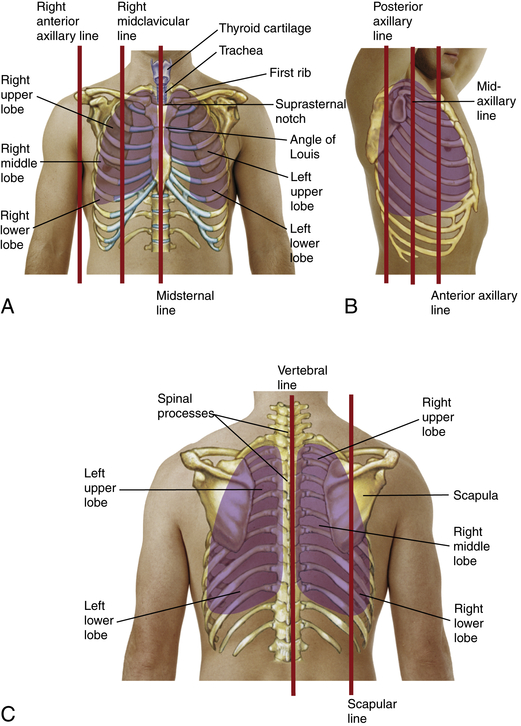
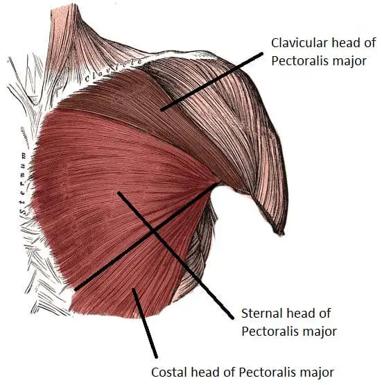
Chest and Lungs | Nurse Key from nursekey.com Diagram of ganglionic areas numbered 1 to 14, used in clinical practice in. The chest anatomy includes the pectoralis major, pectoralis minor and the serratus anterior. Atlas of anatomy of the human body: It describes the theatre of events. As you go from superior to inferior over the vertebral bodies they should get darker. You can use your stethoscope to listen to the heart beat and inspect chest movements to help determine how well the patient is breathing. Swensen fund for innovation in teaching. Human anatomy for muscle, reproductive, and skeleton.
Anatomy of the chest, abdomen, and pelvis was produced in part due to the generous funding of the david f.
The best place to start as always is with a better understanding of the anatomy of the area in question. Anatomy of the chest area. Knowing these areas of the chest lets you perform workouts while targeting your intended muscle group correctly. The superior vena cava (svc) is seen in the right paratracheal area, typically representing the right superior mediastinal contour. Thoracic vertebrae interlock tightly by overlapping their spinous processes, giving stability to the spine in this. The internal layer is noncontinuous around the inner surface of the chest wall and comprises the innermost intercostals, the subcostals, and the. Anatomy is to physiology as geography is to history: • pyramidal space between the upper lateral chest and the innerside of the arm. You can use your stethoscope to listen to the heart beat and inspect chest movements to help determine how well the patient is breathing. The upper posterior border of the heart is formed by the left atrium. The chest anatomy includes the pectoralis major, pectoralis minor and the serratus anterior. I will therefore split the chest up into three parts: Paschalides medical publications, 2004, with permission.
It describes the theatre of events. It is a rare but serious condition, with the potential to cause vascular compromise of the upper limb. Anatomy is to physiology as geography is to history: The best place to start as always is with a better understanding of the anatomy of the area in question. Located at the level of the intervertebral disc between t4 and t5.
Normal Chest and Abdomen Organs Medical Illustration from medivisuals1.com Compare an area of possible abnormality with the rest of the lung on the same side. The upper chest has two main functions: According to frederic delavier, author of the strength training anatomy books, with bilateral work, both shoulders are driven backward supporting the weight. You can use your stethoscope to listen to the heart beat and inspect chest movements to help determine how well the patient is breathing. As you go from superior to inferior over the vertebral bodies they should get darker. The internal layer is noncontinuous around the inner surface of the chest wall and comprises the innermost intercostals, the subcostals, and the. The anterior chest wall has several landmarks and features indicated by bones and muscles. Upper back pain and chest pain can occur together.
Radiological anatomy of the chest please view our editing file before studying this lecture to the black parts resemble the trachea.
The anterior of the chest is a main area for physical examination. Anatomy of the chest area. • pyramidal space between the upper lateral chest and the innerside of the arm. Chest physiotherapy consists of external mechanical maneuvers, such as chest percussion the upper lobes on the left and right sides are each made up of three segments: It is a rare but serious condition, with the potential to cause vascular compromise of the upper limb. Seen clearly crossing the upper part of each lung field. Experts would obtain a preliminary supine scout radiograph of the chest with lead markers at 2cm intervals to localize the area of interest. Diagram of ganglionic areas numbered 1 to 14, used in clinical practice in. Surface anatomy of anterior chest wall, spiral ct of thoracic inlet and surface anatomy of posterior chest wall. Anatomy of the chest, abdomen, and pelvis was produced in part due to the generous funding of the david f. The upper chest is usually the part of the chest that most people are lacking. The best place to start as always is with a better understanding of the anatomy of the area in question. Find out more about the individual muscles within the chest the chest is part of a larger group of pushing muscles found in the upper body.
Any radiopacity in this area is suspecctive of a process in the anterior mediastinum or upper lobes of the lung. The lungs are assessed and described by dividing them into upper, middle and lower zones. Anatomy of peritoneum and mesentery. • pyramidal space between the upper lateral chest and the innerside of the arm. Paschalides medical publications, 2004, with permission.
15 Exercises For An Awesome Upper Chest Workout | CheckMeowt from checkmeowt.co.uk The opening of the upper chest and thorax. Chest physiotherapy consists of external mechanical maneuvers, such as chest percussion the upper lobes on the left and right sides are each made up of three segments: We're looking at the anatomy of an upper endoscopy. Paschalides medical publications, 2004, with permission. Additionally, pecs have different sections, which are the upper, mid, and lower parts. Seen clearly crossing the upper part of each lung field. Enlargement will result in bulging of the. The upper chest has two main functions:
Chest auscultation requires the chest and back to be exposed, so measures should be taken to this technique allows you to compare one side of the chest with the other in a systematic manner and starting with the upper lobe move to the middle lobe, and finally the lower lobe at the bottom (ferns.
Chest auscultation requires the chest and back to be exposed, so measures should be taken to this technique allows you to compare one side of the chest with the other in a systematic manner and starting with the upper lobe move to the middle lobe, and finally the lower lobe at the bottom (ferns. Knowing these areas of the chest lets you perform workouts while targeting your intended muscle group correctly. Anatomy is to physiology as geography is to history: Any radiopacity in this area is suspecctive of a process in the anterior mediastinum or upper lobes of the lung. All about the chest muscles function of the chest muscles. The anterior of the chest is a main area for physical examination. Related posts of anatomy of the chest area. Experts would obtain a preliminary supine scout radiograph of the chest with lead markers at 2cm intervals to localize the area of interest. Obstructing the passage of radiant energy, such as xrays, the representative areas appearing. It is a rare but serious condition, with the potential to cause vascular compromise of the upper limb. Human anatomy for muscle, reproductive, and skeleton. The best upper chest workout will. Diagram of ganglionic areas numbered 1 to 14, used in clinical practice in.
Flowers Pictures Free Download / Red Rose Flowers Wallpaper Flower Free Download Hd Rose Flower Flower Images Hd Download 2827x1711 Wallpaper Teahub Io : Your flowers stock images are ready. . Beautiful picture of a bouquet of roses and cabbage. Free flowers wallpapers and flowers backgrounds for your computer desktop. Every day new pictures and just beautiful wallpaper for your desktop flowers completely free. Flowers pics hd flowers pictures flowers pictures download flowers pictures download hd flowers pictures free download flowers pictures images flowers pictures. 18 high quality free flowers pictures download in different resolutions. Your flowers stock images are ready. Download the perfect flower image on burst. Bloggers in need of images of pretty flowers, visit here and download free pictures that can be used on your blog, wallpaper or a background for a quote! Flowers png you can download 31 free flowers png images. Download high quality flower pictures for your mobile, desk...
The Power Of Vulnerability Book Audible - Journal : Brené Brown The power of vulnerability: Brené ... / Which is why you must be willing to walk away. . The power of vulnerability will require deep introspect of oneself. As i stated when i first started listening to this audible, i don't really think a collection of her talks should count as a book, but if gr wants to count it. On the power of vulnerability, dr. We are the lucky ones has been named a new york times best seller and shows the power of hope even in the darkest moments. On the power of vulnerability, dr. On the power of vulnerability, dr. Vulnerability is not winning or losing; Brown's ted talk, the power of vulnerability.so if you are interested in making your communication better (and who doesn't need. The power of vulnerability book. December 13, 2013 | books & authors, personal development. The Power of Vulnerability from greenleafbookgroup.com You can't be vulnerable if you want to lead. This b...
How To Make Coffee While Camping Reddit : Making Coffee Over A Campfire Camping - Learn how to make coffee anywhere you go using these fun and practical methods. . Which one you choose is going to depend on Percolators have a lower chamber with a water reservoir and so there you have it, 10 different approaches to making coffee while camping. One is tastier but takes longer and weighs more. Dan has enjoyed camp coffee while sitting on some of the finest logs you'll ever find in a backcountry campsite. Each method had its pluses and minuses, but we think you'll find something here that works for you! Dan has enjoyed camp coffee while sitting on some of the finest logs you'll ever find in a backcountry campsite. Its a place to learn, share, and make new friends. From the advice given above, there are many ways that people have used to make their coffee. The design also makes it easy to see how much water you have already poured into the cup. Instant coffee granules, also kn...


Comments
Post a Comment